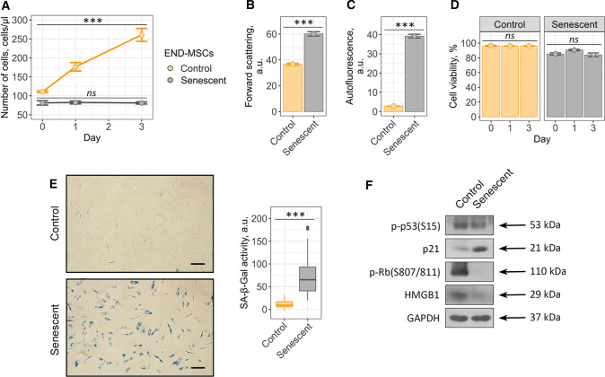
Fig. 1.
Validation of oxidative stress-induced END-MSCs premature senescence model. Senescent END-MSCs a lose proliferation, b undergo hypertrophy, c acquire elevated autofluorescence, retain high cell viability d and e display SA-β-Gal activity as compared to the control ones. f Phosphorylation levels of p53 and Rb and expression levels of p21 and HMGB1 proteins in control and senescent END-MSCs. Values presented are mean ± SD. For multiple groups comparisons at a and d one-way ANOVA was applied, n = 3, ns not significant, ***p < 0.001. For pair comparisons at b, c and e Welch’s t test was used, n = 3 for b and c, n = 50 for e, ***p < 0.001. Scale bars for images are 500 μm. GAPDH was used as loading control

