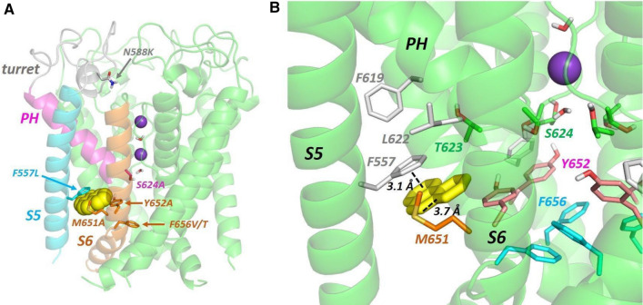
Fig. 6.
A Low energy score pose for phenanthrene (yellow space filling representation) docked into the hERG pore. The hERG subunit with docked phenanthrene shows the location within a single subunit of the mutations described in the text; the structural elements are coloured to match the alignment in Fig. 1a. Purple spheres are K+ ions in the 1 and 3 positions of the selectivity filter. B Close-up of a hydrophobic pocket containing docked phenanthrene (yellow sticks) in the same pose as panel A. An aromatic pi-stacking interaction with the F557 side chain and close interactions with the M651 side chain are indicated with dotted lines

