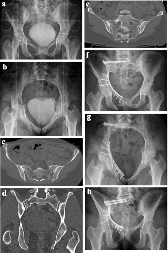
Fig. 4.
a 44-year-old female suffered an unstable pelvic fracture after a fall from 7 m of height. AP-pelvic overview shows displaced superior and inferior pubic rami fractures. b Pelvic inlet view shows a right-sided sacral ala fracture and the right-sided superior and inferior pubic rami fractures. c Axial CT-slice through the posterior pelvis. There is a right-sided sacral fracture. d Coronal CT-slice through the posterior pelvis. The disploaced right.sided sacral fracture is clearly visible. e CT-reconstruction through the longitudinal axis of the sacrum. The sacral fracture runs through the neuroforamina S1 and S2. Dysmorphism of the upper part of the sacrum can be recognized. f Postoperative AP-view of the pelvis. The sacral fracture was stabilized with two iliosacral screws in S1, the superior pubic ramus fracture was stabilized with a buttress plate through the modified Stoppa approach. There were no postoperative problems. No postoperative CT scan was performed. g Pelvic inlet view. h Pelvic outlet view

