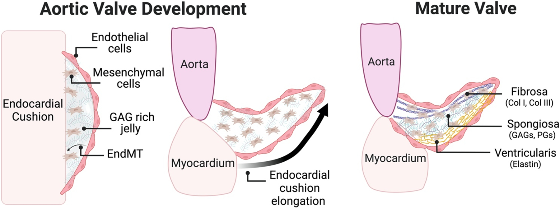
Figure 1: ECM changes during Aortic Valve Development.

In early valve development endothelial cells undergo transition to mesenchymal cells in a GAG-rich jelly. As valvulogenesis continues, endocardial cushions begin to thin and elongate, remaining rich in GAGs. Lastly, the trilaminar structure of the valve begins to emerge in the postnatal period with enrichment of collagen I fibrils in the fibrosa, HA and chondroitin sulfate in the spongiosa, and elastin localized to the ventricularis.
