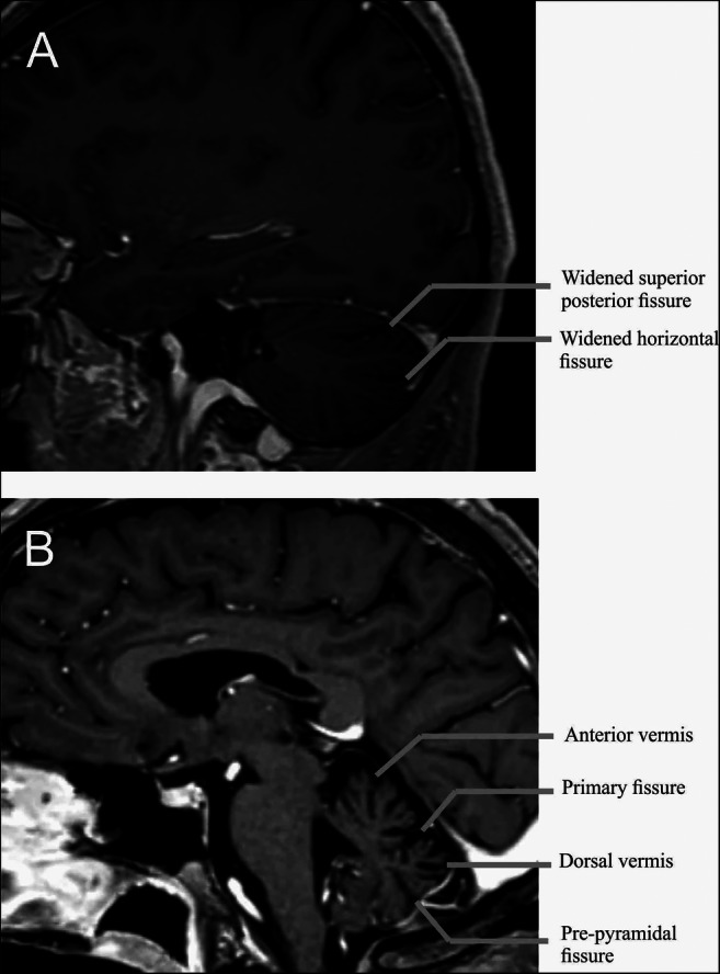
Fig. 1.

Exemple of typical brain MRI in a patient with CANVAS. (A) 3D T1-weighted with gadolinium MRI brain parasagittal section of a 46-year-old CANVAS. This view shows the hemispheric atrophy’s pattern with widening of the superior posterior and of the horizontal fissures, delimiting Crus I. (B) 3D T1-weighted with gadolinium MRI brain midsagittal section of a 70-year-old CANVAS. Note the predominant atrophy of the anterior and dorsal vermis and particularly in the part of the dorsal vermis between the primary fissure and the pre-pyramidal fissure corresponding to the vermal lobules VI, VIIa and VIIb
