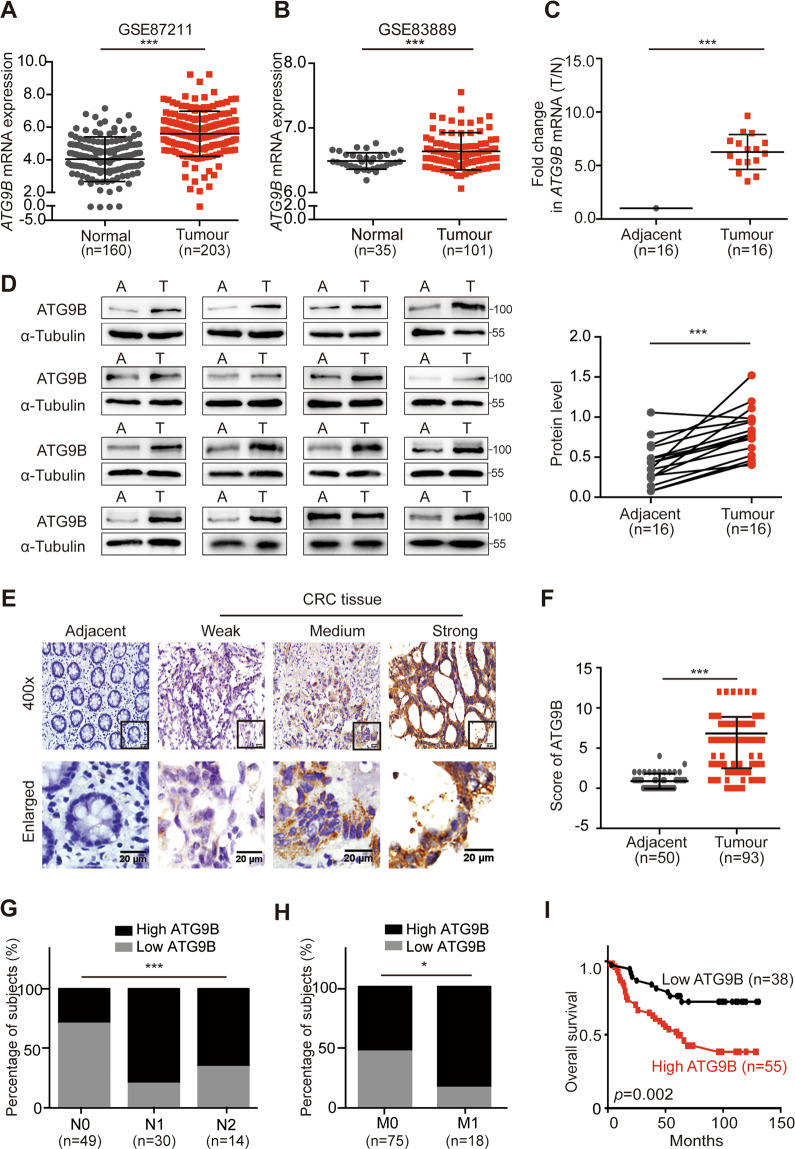
Fig. 1. ATG9B is upregulated in CRC tissues and is positively correlated with poor prognosis in CRC patients.
A, B Analysis of ATG9B expression in CRC tissues compared with adjacent normal tissues in the CRC microarray profiles GES87211 (A) and GSE83889 (B). C Fold change (T/N) of ATG9B mRNA expression in 16 primary CRC tissues and adjacent normal tissues from the same patient, as determined by RT-PCR. D Immunoblot for ATG9B protein expression in 16 human CRC tissues (T) and matched adjacent normal tissues (A) from the same patient. Quantification of protein levels were normalized to those of α-Tubulin are shown in the right panel. E Representative ATG9B immunohistochemical staining images of adjacent normal tissue (Adjacent, n = 50) and tumour tissue (CRC, n = 93) samples (scale bar 20 μm). The magnified parts were displayed in the lower panel. F Immunohistochemical score of ATG9B in adjacent tissues and CRC tumour tissues. G, H Percentage of high and low expression of ATG9B in 93 CRC patients with different N (lymph node metastasis, G) and M (distal metastasis, H) clinical stages. I Kaplan–Meier survival analysis of 93 CRC patients with low and high expression of ATG9B. Data information: Graphs report mean ± SEM. Significance was assessed using two-tailed Student’s t test, except for G, H where Chi-square test was used and I where log-rank test was used. ***P < 0.001, **P < 0.01, *P < 0.05.

