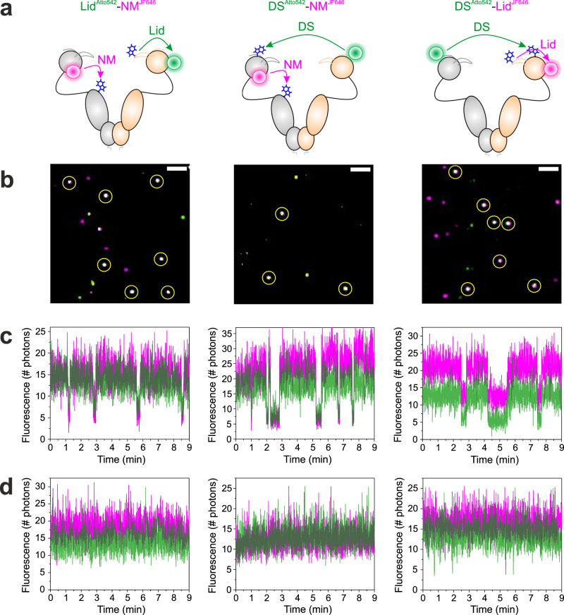
Fig. 4. Two-colour smPET fluorescence microscopy of Hsp90 dynamics.
a Two-colour PET fluorescence reporters on Hsp90 for the simultaneous observation of Lid, DS and NM-association, illustrated as structural models. Fluorophores and Trp are indicated by coloured spheres and blue sticks. The reported conformational changes are indicated by arrows. b Representative two-colour smTIRF images of two-colour PET reporter-containing Hsp90 constructs tethered to glass surfaces (green: Atto542 fluorescence, magenta: JF646 fluorescence). Dually labelled Hsp90 dimers were identified by co-localization of Atto542 and JF646 fluorescence signals (white spots, highlighted by yellow circles). White bars are 1-µm scale bars. c, d Representative two-colour fluorescence intensity time traces recorded from individual Hsp90 molecules containing two-colour PET reporters indicated in panel a. Traces measured in the presence of 4 mM ATP are shown in c. Traces measured in the absence of ATP are shown in d. Panels arranged in vertical columns belong to the same construct (specified in a). Experiments were repeated three times with similar results. Source data are provided as a Source Data file.

