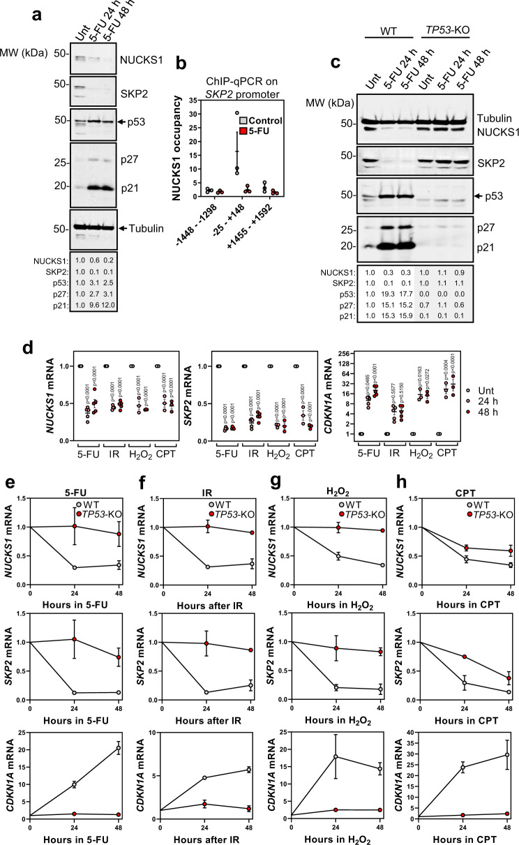
Fig. 5. DNA damage inhibits the NUCKS1-SKP2 axis through p53-mediated transcriptional repression.
a Western blot in WT U2OS cells treated with 50 µM 5-FU for 24 or 48 h. b ChIP-qPCR of NUCKS1 fold enrichment over IgG (=1) on the SKP2 promoter in U2OS cells after treatment with control or 50 µM 5-FU for 48 h. c Western blot in RPE1-hTERT WT and TP53-KO cells treated with 10 µM 5-FU for 24 or 48 h. d RT-qPCR in WT RPE1-hTERT cells treated with 5-FU (10 µM), IR (4 Gy), H2O2 (200 µM), or CPT (100 nM) for 24 or 48 h. Ordinary two-way ANOVA with Dunnett’s multiple comparisons test. e RT-qPCR after 5-FU (10 µM) in RPE1-hTERT WT or TP53-KO cells. f RT-qPCR after IR (4 Gy) in RPE1-hTERT WT or TP53-KO cells. g RT-qPCR after H2O2 (200 µM) in RPE1-hTERT WT or TP53-KO cells. h RT-qPCR after CPT (100 nM) in RPE1-hTERT WT or TP53-KO cells. In a and c, data are representative of 3 independent experiments. In b, d, e, f, g, and h, data are presented as mean ± SEM from 3 (b, h), 3-5 (d) or 2 (e–g) independent experiments. MW: molecular weight, kDa: kilodaltons. Source data are provided as a source data file.

