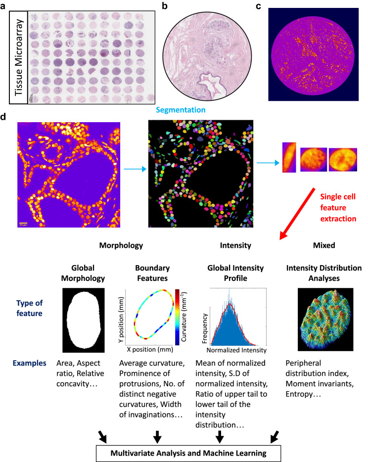
Figure 1.
Imaging based nuclear and chromatin features as single cell biomarkers in tissue biopsies. (a) An overview of the biomax Tissue Microarray (TMA). (b) Representative H&E stained TMA image. (c) Representative Hoescht stained TMA image. (d) The first module of the pipeline automates the thresholding, segmentation and cropping of individual nuclei from large images. A library of individual nuclei is created and passed to the second module where genome architectural (morphological and textural) features are extracted from each nucleus. The consolidated single cell data is then used for further analysis.

