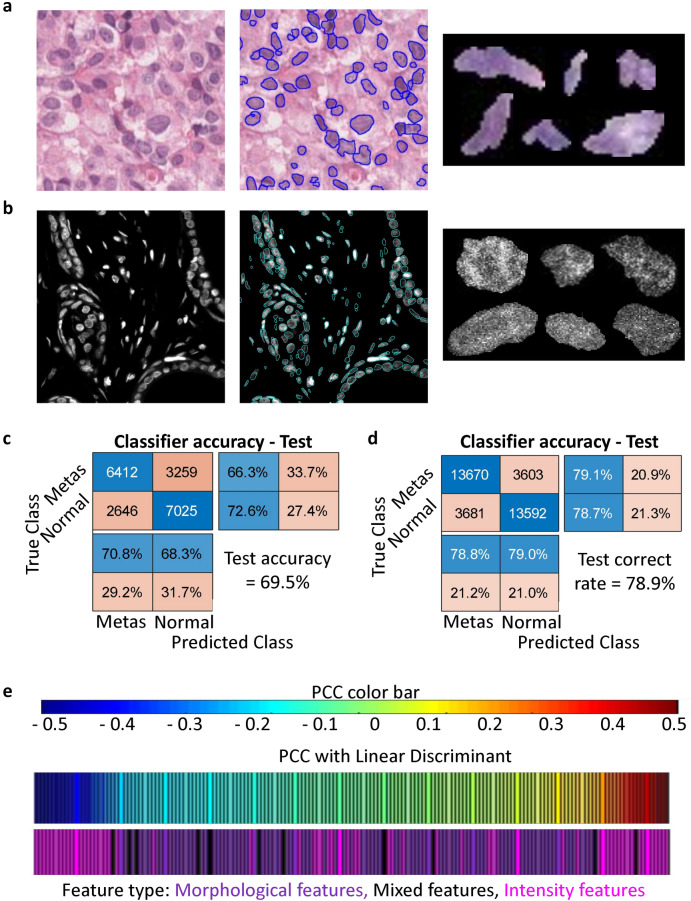
Figure 2.
Hoescht staining improves the sensitivity to discriminate tumor progression. Representative crop of an H&E-stained (a) and Hoescht stained (b) tissue biopsy image (left). Marked in blue are the regions detected as nuclei for downstream segmentation and analysis. Representative crops of individual nuclei segmented are shown on the right side. Confusion matrices depicting the performances of the linear discriminant classifier in classifying nuclei from normal and metastatic tissues according to their nuclear features in H&E-stained tissues (c) and Hoescht stained tissues (d). (e) Correlation between nuclear features and the linear discriminant axis for the Hoescht-stained nuclei. The features have been colour-coded and grouped into morphological features, mixed features and intensity features.

