Figure 5.
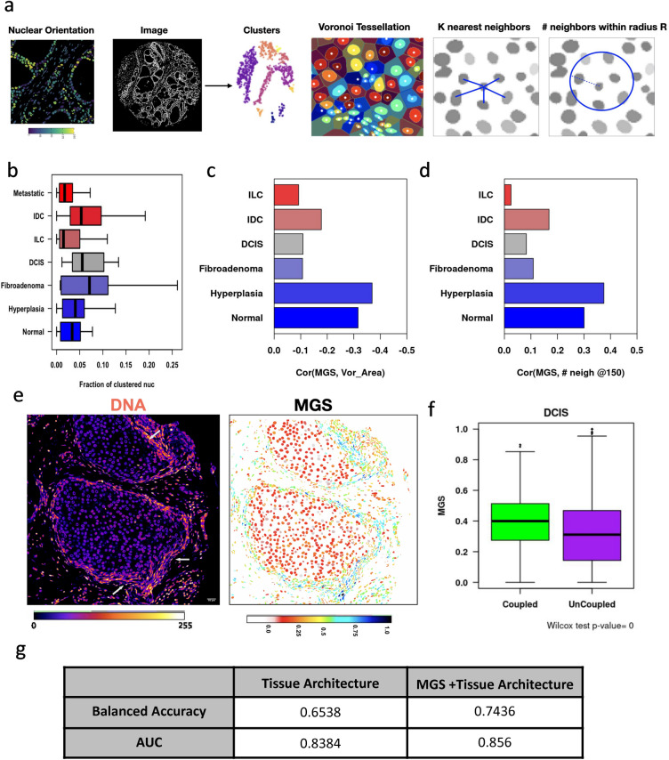
Orientationally coupled regions are characteristic of early breast cancer states. (a) Representation of the method used to identify orientationally coupled regions in the tissue. We first segment nuclei, obtain the angle that the major axis of the fitted ellipsoid makes with the X axis. We filter to obtain only elongated nuclei and then use their centroid and orientation as features for Density-based spatial clustering of applications with noise (DBSCAN). Each identified cluster is depicted in a different color. Local tissue density was calculated using multiple approaches namely, voronoi tessellation, distance to the kth nearest neighbour and number of neighbors in a given radius. (b) Fraction of clustered nuclei in the various stages of breast cancer. One way ANOVA indicated that there were significant changes in the means (p < 0.01). Correlation coefficient between MGS and Voronoi cell area (c) and number of neighbours in 40 µm radius (d). (e) Representative images showing the mechanically coupled regions (arrows) and the MGS of nuclei in Ductal Carcinoma in situ. (f) Mechano-Genomic Score (MGS) of clustered/coupled nuclei (green) and unclustered/uncoupled nuclei (purple) in Ductal Carcinoma In situ (DCIS) TMAs. (g) Tissue level predictions by Linear Discriminant Analysis using Tissue Architecture features with and without Mechano-Genomic Score.

