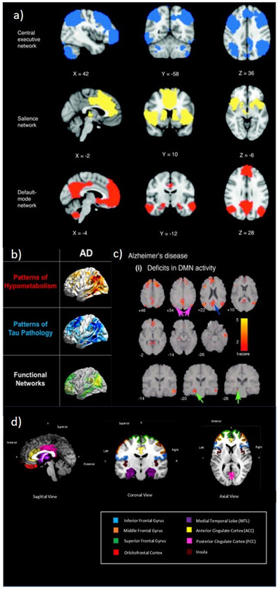
FIGURE 2.

A, Principal brain regions in the central executive, salience, and default‐mode networks (adapted from Menon. 78 B, Overlap between hypometabolism on 18F‐FDG PET, tau aggregation on 18F‐T807 PET, and default mode networks from resting‐state functional MRI in AD (adapted from Drzezga 79 ). C, Changes in DMN activity in patients with AD compared to age‐matched healthy elderly controls. The PCC (blue arrow), angular gyrus in the inferior parietal cortex (magenta arrow) and hippocampus (green arrow) show prominent activity changes in AD (adapted from Menon 78 ). D, Common regions of atrophy and functional impairment associated with deficits in higher level awareness and metacognition in early AD (adapted from Hallam and Huntley 73 ). AD, Alzheimer's disease; DMN, default mode network; FDG, fluorodeoxyglucose; MRI, magnetic resonance imaging; PCC, posterior cingulate cortex; PET, positron emission tomography
