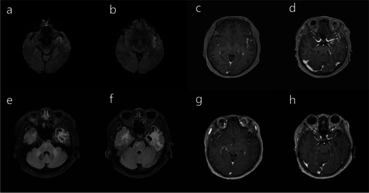
Fig. 1.
a–b Magnetic resonance imaging (MRI) of the brain demonstrated restricted diffusion along the left insular and mesial temporal cortices. c–d Follow-up MRI of the brain performed 1 month after the symptom onset showed contrast enhancement in the corresponding lesions along the left insular and mesial temporal cortices. e–h One month later, MRI demonstrated the subsidence of the contrast enhancement with the encephalomalacic change in the left temporal lobe

