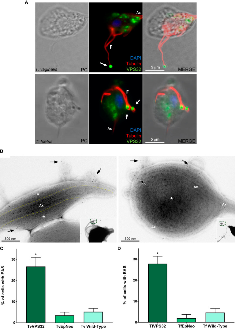
Figure 10.
VPS32 is present in EAS surface, and its overexpression increases EAS formation. (A) Representative immunofluorescence microscopy images of Trichomonas vaginalis and Tritrichomonas foetus exogenously expressing TvVPS32 and TfVPS32 with a C-terminal hemagglutinin (HA) tag, respectively, using a rabbit anti-HA antibody (green). PC, phase-contrast image. The flagella (F) and axostyle (As) are labeled with mouse anti-tubulin antibody (red). Arrows indicate the subcellular localization of VPS32 in structures similar to flagellar swelling at flagella tip. The nucleus (blue) is stained with 4′,6′-diamidino-2-phenylindole (DAPI). The cytosolic subcellular localization of VPS32 protein is also noticed. (B) Negative staining of TvVPS32-HA-transfected parasites immunogold-labeled with anti-HA antibody demonstrates that TvVPS32 is localized in the surface of extra-axonemal structures (EASs) as well as in MVs that protrude from EASs (arrows). (C–D) Analysis of the percentage of EASs in the flagella of TvVPS32FL (C) and TfVPA32FL (D) parasites. Three independent experiments in duplicate were performed, and 100 parasites exhibiting at least one swelling were randomly counted per sample using a phase-contrast microscope. Data are expressed as means ± SD. Approximately 27% and 28% of flagellar EASs were observed in TvVPS32- and TfVPS32-transfected parasites, respectively, compared with 2%–5% of EASs observed in EpNeo (empty plasmid transfected) and wild-type parasites. *p < 0.05 compared with EpNeo and wild-type parasites using one-way ANOVA test (Kruskal–Wallis test; Dunn’s multiple comparisons test).

