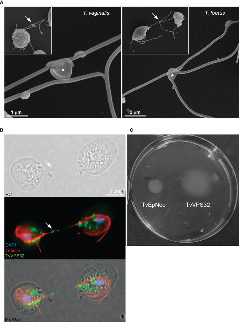
Figure 11.
TvVPS32 might play a role in parasite motility. (A) Representative SEM images of parasites (Trichomonas vaginalis and Tritrichomonas foetus) connected to themselves by EASs (arrows). Notice the EASs in higher magnification (*). (B) Immunofluorescence images showing that TvVPS32-transfected parasites connect with each other through the flagella and that TvVPS32 is localized in the flagella of parasites in contact. TvVPS32 parasites cultured in the absence of host cells were co-stained with anti-HA (green) and tubulin (red). The nucleus (blue) was also stained with DAPI. Arrows indicate the EASs. PC, phase-contrast image. The cytosolic subcellular localization of VPS32 protein is also noticed. (C) Representative TvVPS32 parasite motility assay. TvEpNeo (empty plasmid transfected) and TvVPS32 parasites were spotted onto soft agar, and their migration capacity was analyzed by measuring the size of the halo diameter during 4 days under microaerophilic conditions at 37°C. TvVPS32 parasites showed a higher capacity of migration compared with TvEpNeo parasites.

