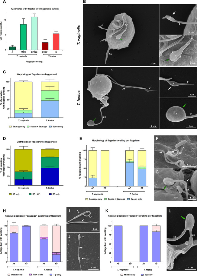
Figure 2.
Morphological analyses of flagellar swellings in Trichomonas vaginalis and Tritrichomonas foetus under standard growth conditions. (A) Quantification of the percentage of parasites that display flagellar swellings. The values are expressed as the means ± standard deviation (SD) of three independent experiments, each performed in duplicate. Five hundred parasites per sample were randomly counted. (B) General and detailed views of flagellar swellings (*) in T. vaginalis and T. foetus obtained by SEM. The swellings can exhibit two different morphologies: “sausage-shaped” (white arrows) and “spoon-shaped” (green arrows). Notice that the “sausage-like” swelling runs laterally to the flagellum (F), whereas in the spoon-shaped structure, the swelling is surrounded by the flagellum. AF, anterior flagella; RF, recurrent flagellum; As, axostyle. (C, D) Quantitative analysis of the morphology (C) and distribution (D) of flagellar swellings per parasite. Three independent experiments in duplicate were performed, and 100 parasites exhibiting at least one swelling were randomly counted per sample using SEM. Data are expressed as percentage of parasites with flagellar swelling ± SD. AF, anterior flagella; RF, recurrent flagellum. (E) Quantification of the morphology of flagellar swelling per flagellum. The values are expressed as the means of the percentage of flagellum with swelling ± SD of three independent experiments, each performed in duplicate. One hundred anterior and recurrent flagella with swelling per sample were randomly counted using SEM. AF, anterior flagella; RF, recurrent flagellum. (F, G) Detailed views of RF of T. vaginalis (F) and AF of T. foetus (G) by SEM. UM, undulating membrane. In (F), a sausage-shaped swelling (arrow) is seen at the tip of the flagellum. Notice in (G) the presence of “sausage” (white arrow) and “spoon-like” (green arrow) structures in the same flagellum. (H) Analysis of the relative position of “sausage” swelling per flagellum. Three independent experiments in duplicate were performed, and 100 anterior and recurrent flagella with swelling per sample were randomly counted using SEM. Data are expressed as percentage of flagellum exhibiting swelling ± SD. AF, anterior flagella; RF, recurrent flagellum. (I, J) SEM of sausage-shaped structures (arrows) located along the AF of T. vaginalis (I) and at the tip and in the middle of the same recurrent flagellum of T. foetus (J). (K) Quantification of the relative position of “spoon” swelling per flagellum. The values are expressed as the means of the percentage of flagellum exhibiting swelling ± SD of three independent experiments, each performed in duplicate. One hundred anterior and recurrent flagella with swelling per sample were randomly counted using SEM. AF, anterior flagella; RF, recurrent flagellum. (L) SEM of a spoon-shaped structure (arrow) located in the middle of T. foetus RF. UM, undulating membrane.

