Figure 7.
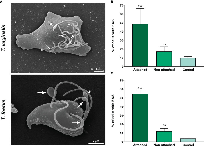
The EAS formation increases during trichomonad attachment on fibronectin-coated coverslips. (A) SEM of Trichomonas vaginalis and Tritrichomonas foetus after adhesion assay on fibronectin-coated coverslips. Arrows indicate the EAS. Notice that parasites display an amoeboid morphology. (B, C) Quantitative analyses in T. vaginalis (B) and T. foetus (C). The percentage of cells with EASs was determined by counting 500 parasites per sample using SEM. Data are expressed as means of three independent experiments in duplicate ± SD. Attached and non-attached: parasites resuspended in PBS incubated on fibronectin-coated coverslips in a humidity chamber for 2 h at 37°C and rigorously washed with PBS to remove non-attached cells. Attached parasites remain on the coverslips even after several washes. Non-attached parasites were collected with a pipette, harvested by centrifugation, and prepared for SEM. Control, parasites incubated on uncoated coverslips under the same conditions mentioned above, collected with a pipette, harvested by centrifugation, and prepared for SEM. “Control” is formed by non-adherent, suspended cells from uncovered coverslips, whereas non-adherent parasites from fibronectin are called “non-attached.” The percentage of parasites displaying EASs is significantly higher in the attached group when compared with the non-attached and control groups. ***p < 0.001 compared with the control group using one-way ANOVA test (Kruskal–Wallis test; Dunn’s multiple comparisons test). ns, non-significant.

