Figure 8.
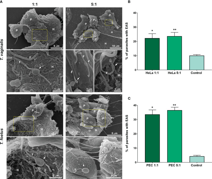
EASs are formed in response to host cell exposure. (A) Representative SEM images of Trichomonas vaginalis and Tritrichomonas foetus after host cell interaction. HeLa and bovine preputial epithelial cells (PECs) were co-incubated with T. vaginalis and T. foetus, respectively, at cell ratios of 1:1 or 5:1 parasite:host cell in PBS-F (PBS with 1% FBS at pH 6.5) at 37°C for 30 min. Flagellar swelling (arrows) are seen in some parasites (P). Notice that some swellings are in direct contact to the host cells (H). (B, C) Quantification of the percentage of T. vaginalis (B) and T. foetus (C) with flagellar swelling after the host cell interaction. Three independent experiments in duplicate were performed, and 500 parasites were randomly counted per sample using SEM. Data are expressed as percentage of parasites ± SD. For the control experiments, parasites incubated in PBS in the absence of host cells were analyzed. The percentage of parasites with flagellar swelling increases after the hot cell exposure when compared with control (PBS). *p < 0.05; **p < 0.01 compared with control using one-way ANOVA test (Kruskal–Wallis test; Dunn’s multiple comparisons test).

