Figure 9.
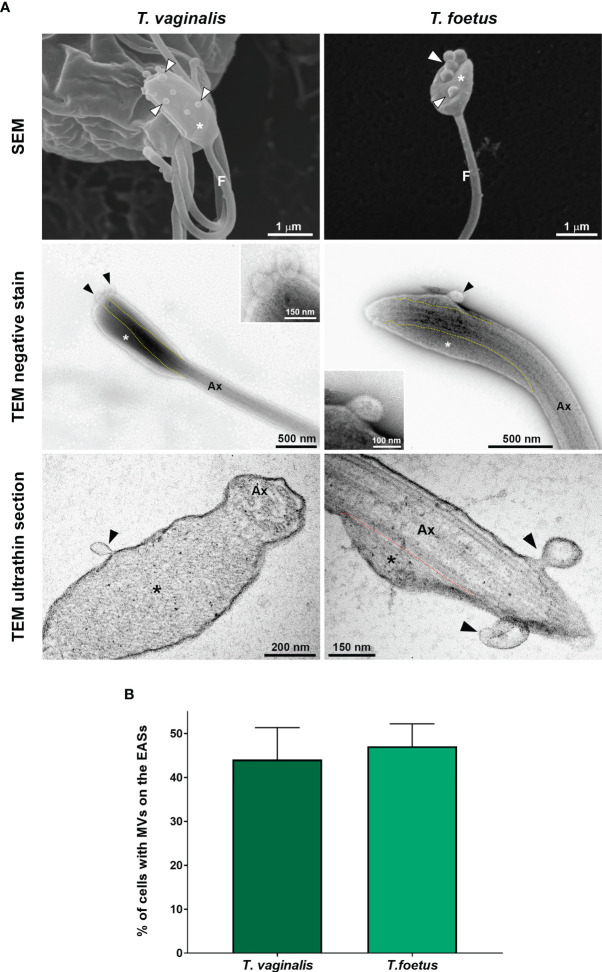
EASs release microvesicle-like structures. (A) Representative micrographs of MVs (arrowheads) protruding from the flagellar membrane of the EASs (*) of Trichomonas vaginalis and Tritrichomonas foetus. The images were obtained by SEM (first row), negative staining (second row), and ultrathin sections (third row). The dotted lines indicate the boundary between axoneme (Ax) and the extra-axonemal filaments (*). (B) Percentage of EASs with protruding MVs on their surface. Three independent experiments in duplicate were performed, and 100 parasites exhibiting at least one swelling were randomly counted per sample using SEM. Data are expressed as means ± SD. Approximately 45% of parasites with flagellar swelling exhibited associated MVs.

