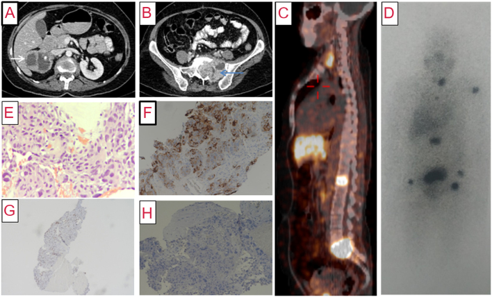
Figure 2.
(A) Contrast-enhanced computed tomography (axial section) of abdomen showing a predominantly cystic mass lesion (6.1 × 5.5 × 5.5 cm) in the right suprarenal region (white arrow) with peripherally enhancing solid component. (B) Caudal section in the same scan showing a large lytic lesion with soft tissue (7.3 × 5.9 × 6.8 cm) in the sacral body and the left ala (blue arrow), with similar enhancement characteristics. (C) 68Ga-DOTATATE PET-CT showing somatostatin receptor avid lesions in thyroid, L1 vertebral body, and sacrum. (D) 131I-MIBG scan (anterior view) showing areas of increased radiotracer uptake in the thyroid bed, left humerus, L1 vertebra, sacrum, and both pelvic bones (black). (E) Photomicrograph of biopsy of sacral mass showing metastatic pheochromocytoma with nuclear pleomorphism and moderate to abundant amphophilic cytoplasm with perivascular arrangement of tumor cells (×200, hematoxylin and eosin). (F) Tumor cells showing positive staining for chromogranin immunohistochemistry (IHC), suggesting neuroendocrine tumor (×100, 3,3'-diaminobenzidine (DAB)). (G) Tumor cells showing nuclear reactivity for GATA 3 IHC, favoring pheochromocytoma (×40, DAB). (H) Tumor cells showing negative staining for calcitonin IHC, ruling out medullary thyroid carcinoma (×100, DAB).

 This work is licensed under a