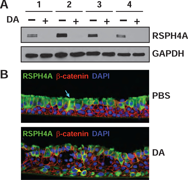
Figure 3. The cilia-specific marker, RSPH4A was markedly reduced after DA treatment.
A) The levels of RSPH4A and GAPDH in cell lysates were visualized by western blotting. Western blotting performed on a replicate across the entire MW range (Figure S2) showed that only a single antigen was recognized by this antibody. B) Immunofluorescence was performed on cross sections of formalin-fixed and paraffin-imbedded TBECs to visualize RSPH4A (green, Alexa Fluor 488), β-catenin (red, Alexa Fluor 594) and nuclei (blue, DAPI) in donor 3. Localization of RSPH4A in ciliated cells is shown by blue arrow (upper image) and localization to basal cells is shown by yellow arrow (lower image). Images in (B) are representative of three different sections per condition (Fig. S3, A–B), and results were replicated in single sections from donors 2 and 4 (Fig. S3, C–F).

