
FIGURE 1.
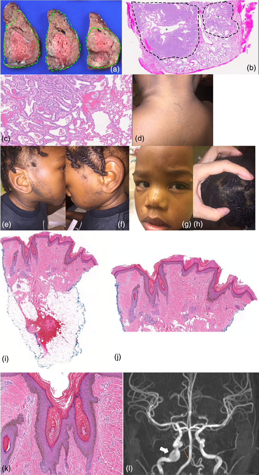
Clinical and pathologic patient features. (a–c) CPAM. (a) Gross pathology shows a solid, lobulated mass occupying the majority of the lobe of lung (outlined in green). The peripheral, better-fixed part of the lesion is tan-brown while the center unfixed area is red. There are multiple central branching airway structures without prominent cystic change. (b) The low power view of a lobule shows a central branching airway-like structure (*) surrounded by malformed parenchyma with increased mesenchyme (outlined by dotted line) with peripheral malformed lung with a more mature appearance (H&E, 0.8×). (c) High power view shows the junction between the two areas with low columnar epithelium lining abundant mesenchyme in septa between small airspaces (left) and (d) more mature but still enlarged alveoli with decreased mesenchyme and flattened epithelium (right; H&E, 13.4×). (e,f) Beard like distribution of comedones. (g) Comedones at midline face. (h) Area of wooly hair. (i–k) Nevus comedonicus biopsy specimen. (i) Scanning view of this punch biopsy specimen demonstrates a sparse inflammatory infiltrate, and the epidermis shows hyperkeratosis and papillomatosis. There are multiple areas consistent with comedones, with small cystic areas that contain hyperkeratosis. The focal hemorrhage in the adipose tissue is related to the surgical procedure to obtain the specimen (H&E, 20×). (j) Medium power view demonstrates the epidermal changes consistent with an epidermal nevus, with epidermal papillomatosis and hyperkeratosis. The areas consistent with comedones are also shown, with cystic areas containing hyperkeratotic keratin (H&E, 50×). (k) This high power view demonstrates an area of the specimen with features of comedones. There are areas of cystic epithelium which contain hyperkeratotic keratin (H&E, 100×). (l) Brain MRI demonstrating a 10 mm aneurysm of the right internal carotid artery lacerum segment (white arrow) and absent right posterior inferior cerebellar artery (small orange arrow) [Color figure can be viewed at wileyonlinelibrary.com]