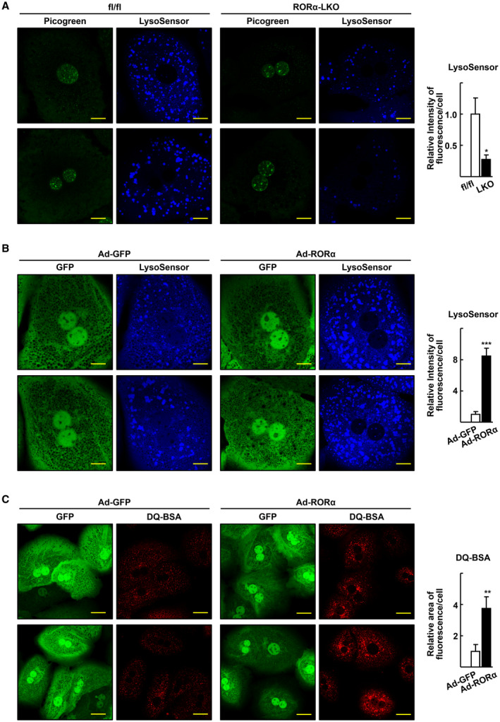
FIG. 1.

RORα enhances lysosomal acidification in the hepatocytes. (A) Confocal microscopy was performed with primary hepatocytes obtained from the RORαfl/fl (fl/fl) and RORα‐LKO (LKO) and stained using LysoSensor Blue DND‐167. Picogreen was used to stain the nucleus. Representative images of hepatocytes are presented. Fluorescence intensity was quantified in at least 100 cells using ImageJ. Scale bar: 10 μm. *P < 0.05 versus fl/fl. (B) After mouse primary hepatocytes were infected by either Ad‐GFP or Ad‐RORα for 18 hours, cells were stained with LysoSensor Blue DND‐167 and subjected to confocal microscopy. Representative images of hepatocytes are presented. Fluorescence intensity was quantified in at least 100 cells using ImageJ software. Scale bar: 10 μm. ***P < 0.001 versus Ad‐GFP‐infused hepatocytes. (C) The virus‐infused hepatocytes were incubated with DQ Red BSA 20 μg/mL for 16 hours and subjected to confocal microscopy. Representative images are shown. Fluorescent area was quantified in at least 100 cells using ImageJ software. Scale bar: 25 μm. **P < 0.01 versus Ad‐GFP.
