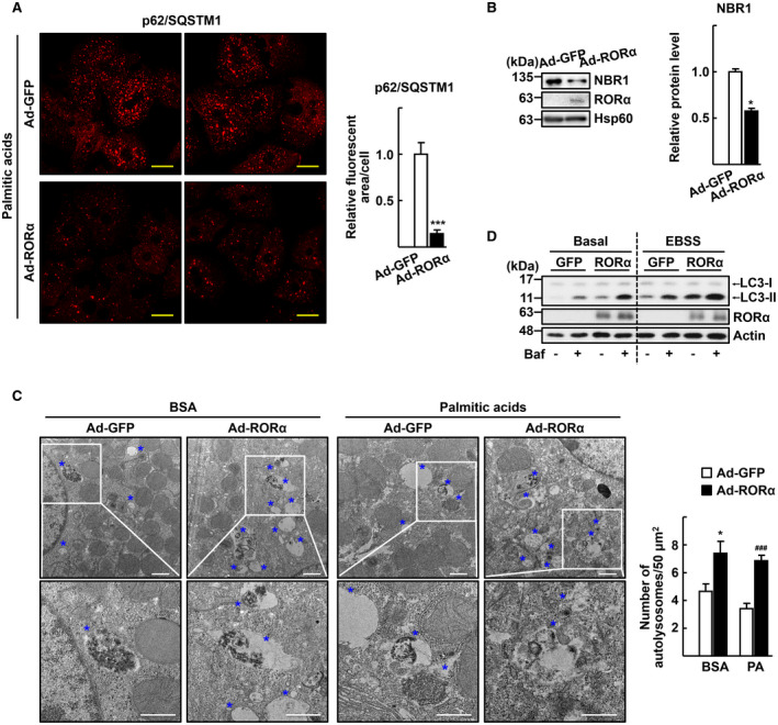
FIG. 5.

Infusion of Ad‐RORα enhances autophagic flux in the hepatocytes. Primary hepatocytes obtained from C57/BL6 mice were infected by either Ad‐GFP or Ad‐RORα for 18 hours. (A) The virus‐infused hepatocytes were exposed to 0.5 mM palmitic acids conjugated with BSA for 1 hour. Immunostaining was performed for p62/SQSTM1 (red) and examined by confocal microscopy. Representative images are shown. The relative area of fluorescence was quantified using Image J software in at least 100 cells. Scale bar: 25 μm. ***P < 0.001 versus Ad‐GFP. (B) The virus infused hepatocytes were exposed to 0.5 mM palmitic acids conjugated with BSA for 1 hour. Expression of the NBR1 protein was analyzed by western blotting. Band intensity of NBR1 was quantified using ImageJ software and normalized to that of Hsp60. *P < 0.05 versus Ad‐GFP (n = 4). (C) The virus‐infused hepatocytes were exposed to 0.5 mM palmitic acids conjugated with BSA for 1 hour and then subjected to EM. Representative EM images with blue stars indicating autolysosomes are shown. The number of autolysosomes was counted in at least 20 cells. Scale bar: 1 μm. *P < 0.05 versus Ad‐GFP with BSA and ### P < 0.001 versus Ad‐GFP with palmitic acid BSA. (D) The virus‐infused hepatocytes were exposed to Earle’s balanced salt solution for 1 hour with or without 30 nM bafilomycin A1 for examination of autophagy flux. Accumulation of LC3‐II protein was analyzed by western blotting.
