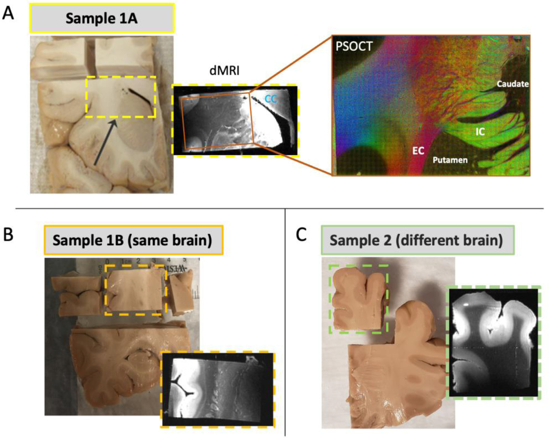
Fig. 1.

Overview of sample identification and data acquisition. Ex vivo human brain samples were extracted from coronal slabs (dashed rectangles) and dMRI data were acquired at 9.4T. Single slices from b=0 scans are shown for each sample. Samples 1A and 1B were cut from different anatomical locations of the same brain, and sample 2 was extracted from a different brain. Following dMRI, a piece of sample 1A was cut and imaged with PSOCT (A, right). Sample 1A, which contained the corpus callosum (CC), internal capsule (IC), external capsule (EC), caudate and putamen, was used as the test dataset. Each of samples 1A, 1B, and 2 were used as training datasets.
