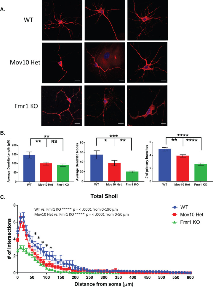
Fig 1. Mov10 Het hippocampal neurons show an abnormal morphology similar to Fmr1 KO neurons.
(A) MAP2/DAPI immunostaining of hippocampal neurons from DIV14 WT, Mov10 het, and Fmr1 KO neurons. Neurons were prepared from 3 independent litters of each genotype and thus, 3 independent cultures. The total number, N, was compiled from the three biological experiments. (B) Dendritic morphology analysis of average dendritic length, dendritic nodes and primary branches. Confocal z-stacks of MAP2-stained WT, Mov10 het and Fmr1 KO DIV14 neurons were analyzed. (C) Dendritic morphology analysis. Confocal z-stacks of MAP2-stained WT, Mov10 het and Fmr1 KO DIV14 neurons were analyzed using Sholl. Statistics were calculated using two-way ANOVA followed by Bonferroni multiple comparisons test. Error bars indicate SEM and *p < 0.05; ****p < 0.0001 (n = 56 neurons for WT, n = 94 neurons for Mov10 Het, n = 58 for Fmr1 KO). Scale bar = 10 μm.

