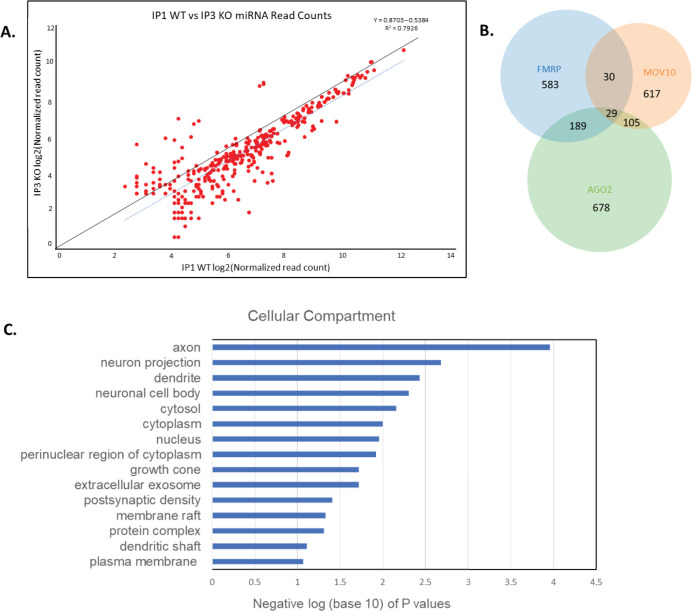
Fig 4. Global miRNA reduction in brain in the absence of FMRP.
(A). Two-fold less miRNAs associate with AGO2 in the absence of FMRP. Reads per million of WT (X-axis) and Fmr1 KO (Y-axis) AGO2-IPs at each cluster that maps to miRNAs. The IPs had a log2 fold change ≥3 over input and p-value ≤ 0.001. Solid black line = best fit of data. Dashed blue line = actual fit of data. (B). Venn diagram showing the overlap between brain-derived iCLIP targets of FMRP [2], MOV10 [15], and AGO2. All three proteins in the brain commonly bound 29 mRNAs (Dicer1 included). (C). GO analysis of the shared mRNAs from postnatal brain. Y axis: GO terms for Cellular Compartment; X axis: negative log (base 10) of the 15 lowest p values showing FMRP binds mRNAs encoding proteins involved in neuron projection.

