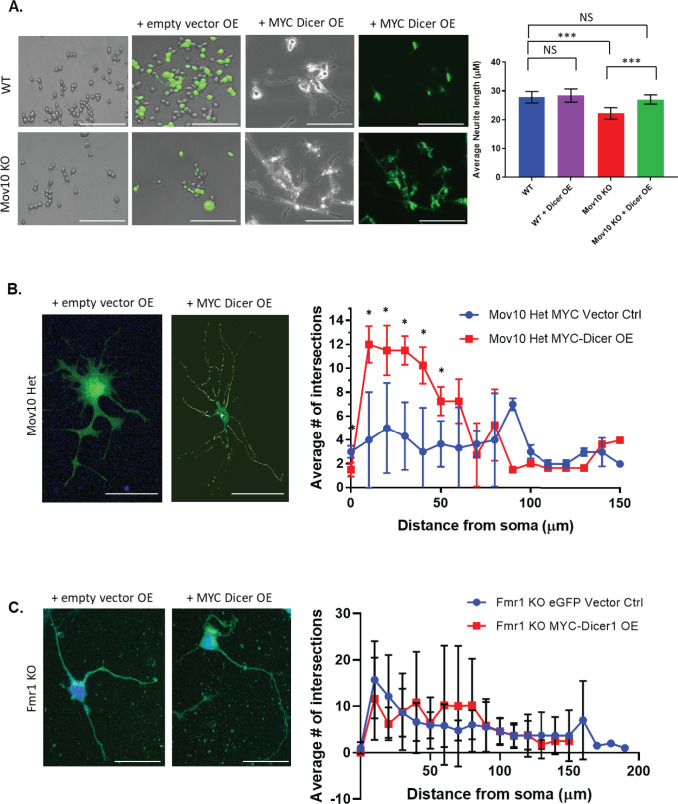
Fig 7. Overexpression of MYC-Dicer1 rescues impaired neuronal phenotype.
(A) Brightfield and immunofluorescence images of WT or Mov10 ko N2A, untransfected or transfected with the empty MYC-vector or the MYC-tagged Human Dicer1, as indicated across the top, and stained with the anti-MYC antibody. The average neurite length was measured and shown on the right. Scale bar = 200 μm. Neurite length in micrometers was measured as described in the Methods. All measured data are expressed as means ± SEM. ***p < 0.001 (Student’s t-test with Welch’s correction). (B,C). Empty vectors, either MYC or eGFP, and MYC-tagged Human Dicer1 was transfected (over-expressed [OE]) in Mov10 HET hippocampal neurons (B) and Fmr1 KO hippocampal neurons (C) at DIV 2 followed by immunofluorescence for MYC at DIV 7. Sholl statistics were calculated using two-way ANOVA followed by Bonferroni multiple comparisons test. Error bars indicate SEM and *p < 0.05 (n = 3 neurons for Mov10 Het empty MYC-vector control, n = 5 neurons for Mov10 Het MYC Dicer overexpression, n = 5 for Fmr1 KO MYC Dicer overexpression, n = 5 for eGFP vector control. Bubbles were removed from Mov10 Het OE images for easier viewing. Scale bar = 100 μm.

