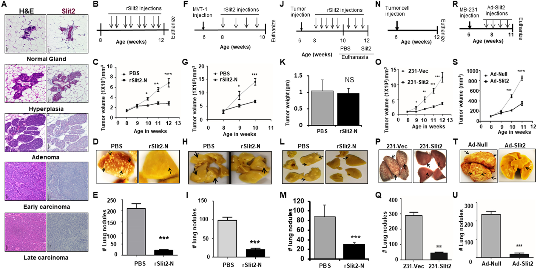
Figure 1: rSlit-N treatment inhibits breast cancer growth and metastases.

(A) Mammary glands were isolated from MMTV-PyMT at different stages of tumor progression and tissue sections were stained with H&E stain or immunostained with Slit2 antibody using the IHC technique.
B-E. (B) Eight weeks old MMTV-PyMT mice bearing spontaneous mammary tumors were treated with rSlit2-N or PBS for four weeks. (C) Tumor volume was measured every week up to 12 weeks of age. (D) Representative pictures of the lungs harvested from (B). (E) The number of metastatic nodules in the lungs.
F-M. MVT1 cells were orthotopically implanted into the six weeks old FVB wild type mice. (F) Schematics of mice treatment with rSlit2-N or PBS. N=6 mice in each group. (G) Tumor volume was measured every week. (H) Representative pictures of the lungs harvested from (F). (I) The number of metastatic nodules in the lungs. (J) Schematics of mice treatment with rSlit2-N or PBS, showing Slit2 treated tumors were allowed to grow for additional two weeks. N=5 mice in each group. (K) At the end, tumors were harvested and weight was measured. (L) Representative pictures of the lungs harvested from (J). (M) The number of metastatic nodules in the lungs.
N-Q. (N) Human breast cancer cell line MDA-MB-231 overexpressing Slit2 (231-Slit2) or vector control (231-Vec) implanted orthotopically into the NSG mice. (O) After two weeks of tumor injection, tumor volume was measured every week up to the age of 12 weeks. (P) The images of lungs harvested from (N). (Q) The harvested lungs were analyzed for the number of metastatic nodules using a dissection microscope.
R-U. NSG females were injected with MDA-MB-231 cells and treated with Adeno-Slit2 or Adeno-Null. (R) Schematics of mice treatment with Adeno-Slit2 or Adeno-Null. (S) Starting from 8 weeks of age, tumor volume was measured every week. (T) Representative pictures of the lungs harvested from (R). (U) The number of metastatic nodules in the lungs. * is p<0.05, ** is p<0.01, *** is p<0.001, NS is P value not significant using student’s t-test.
