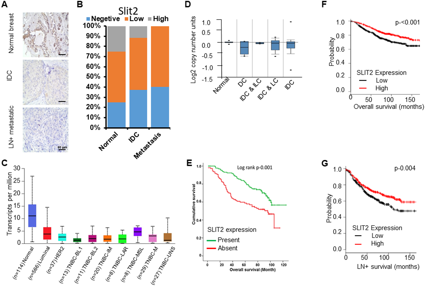
Figure 6. Slit2 expression levels predict breast cancer patient survival.

(A) Human breast tissue microarray (pilot TMA) was immunostained with Slit2 antibody using IHC. Representative images showing Slit2 expression levels in normal (n=10), infiltrative ductal carcinoma (IDC) (n=50) and lymph node-positive metastatic (LN+ metastatic) (n=40) breast tissues.
(B) The graph depicts the percentage of human breast samples expressing different levels of Slit2.
(C) The expression level of Slit2 in human breast tissues using UALCAN database. BL1, basal-like 1; BL2, basal-like 2; IM, immunomodulatory; M, mesenchymal; MSL, mesenchymal stem-like; LAR, luminal androgen receptor; UNS, unspecified.
(D) Analysis of Slit2 gene copy number in TCGA dataset using Oncomine. N, normal breast (n-111); DC, ductal carcinoma (n-5); IDC & ILC, invasive ductal and invasive lobular carcinoma (n-5); IDC & LC, invasive ductal and lobular carcinoma (n-14); IDC, invasive ductal carcinoma (n-639).
(E) Using experimental TMA, the association of Slit2 protein expression levels with patient survival was analyzed. The Kaplan-Meier graph demonstrating the association of Slit2 expression level with patient survival.
(F) Analysis of breast cancer patient overall survival based on expression levels of Slit2 mRNA in the KM-plotter database (n=1115).
(G) Analysis of breast cancer lymph node-positive (LN+) metastases patient survival based on expression levels of Slit2 in the KM-plotter database (n=936).
