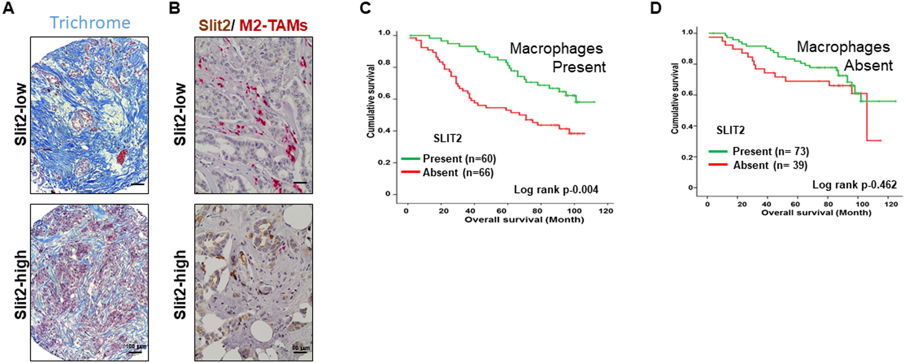
Figure 7. Slit2 expression inversely correlates with TAMs and fibrosis in the breast cancer patient.

(A) The images showing the detection of fibrosis in patient samples by Trichrome staining.
(B) The representative images of breast cancer patient sample immunostained using antibodies specific for Slit2 and CD163 and double color IHC.
C-D. The TMA was stained with macrophage-specific CD68 antibody and patients were divided into macrophage present or macrophage absent groups. (C) The graph showing the survival analysis of patients with macrophages based on Slit2 expression present or absent. (D) The graph showing the survival analysis of patients without macrophages based on Slit2 expression present or absent.
