Figure 3.
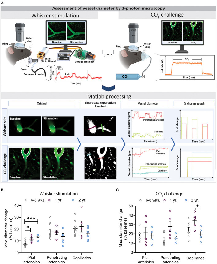
Assessment of vessel diameter by 2-photon microscopy. (A) Experimental setup for whisker stimulation (top left) and hypercapnia during (top right) and a schematic drawing of the data analysis work flow by the newly developed Matlab script. (B) Quantification of maximal diameter changes in different vessel segments at different ages following whisker stimulation. Pial vessel reactivity increased with age (*P < 0.05: 6–8 weeks vs. 1 year and ***P < 0.001: 6–8 week vs. 2 year) while over all the capillary response was reduced in 2-year-old mice in comparison to young and 1-year-old mice. (C) Quantification of maximal diameter changes in different vessel segments at different ages following hypercapnia. Neurovascular reactivity of cerebral capillaries was impaired in 2-year-old mice as compared to 6–8 weeks and 1-year-old mice (*P < 0.05: 1 year vs. 2 year) (mean ± SEM, n = 4–10 mice/group, One-way ANOVA).

