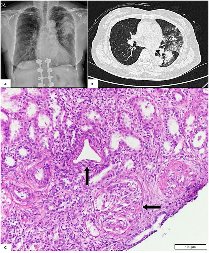
Figure 1.
(A) Pulmonary X-ray showing dense patchy consolidations in the left upper lung field. (B) Pulmonary computed tomography revealing peribronchial consolidations and ground glass opacities in the bilateral lung field, predominantly in the left upper lobe. (C) Light microscopy showing predominant vasculitis (parallel arrow) with pauci-immune crescent glomerulonephritis (horizontal arrow).

