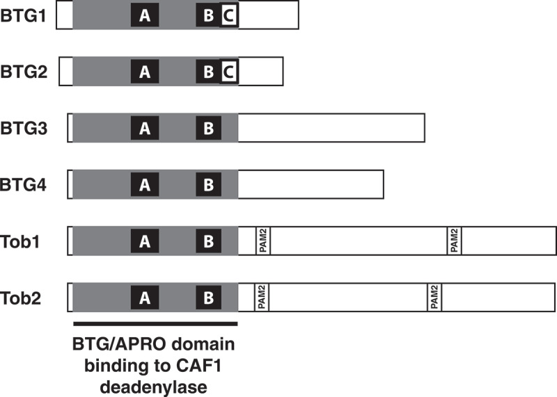
Figure 1.

Organization of BTG/Tob proteins
The six human BTG/Tob proteins are schematically presented to scale with grey box indicating the APRO domain that binds to the CAF1 deadenylase. This domain contains the conserved A and B boxes as well as, in the case of BTG1 and BTG2 the C box. The scheme also indicates the location of PAM2 motifs which are present in the C-terminal extensions of Tob1 and Tob2. The latter interact with the MLLE domain of PABPC proteins.
