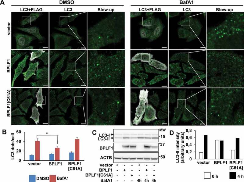
Figure 4.

BPLF1 alters the autophagic flux. (A) Endogenous LC3 was detected by immunofluorescence in Hela cells transfected with either empty vector, BPLF1 or BPLF1[C61A]. Cells expressing catalytically active BPLF1 showed a small increase of LC3 puncta at steady state and significantly decreased accumulation of LC3 puncta upon treatment with BafA1. Scale bar: 10 μm. (B) Quantification of data presented in A; mean ± SEM of data pooled from three independent experiments. Statistical analysis was performed using Student t-test. *P ≤ 0.05. (C) Western blot illustrating the increase of lipidated LC3-II in cells expressing catalytically active BPLF1 and failure to accumulate LC3-II upon treatment with BafA1. (D) The intensity of the LC3-II bands was quantified by densitometry
