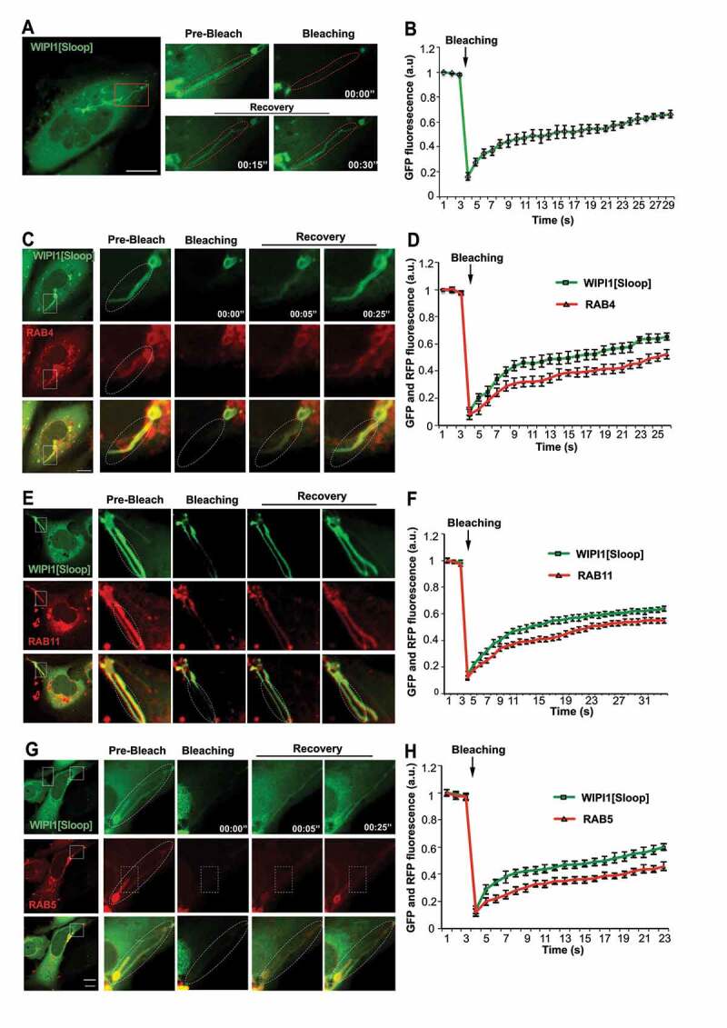
Figure 9.

WIPI1-tubules are continuous with endosomes. (A) In HK2 cells expressing EGFP-WIPI1[Sloop], EGFP-labeled tubules (in the boxed area) were photobleached. EGFP fluorescence is shown before and at different times after bleaching. The dashed line indicates the bleached region. The boxed area is shown at higher magnification. Scale bar: 10 μm (B) Quantification of experiments as in a. Means ± s.d. are shown. Data are representative of 3 independent experiments with at least 20 cells analyzed in each experiment. C-H. Experiments as in (A) and (B), respectively, but with cells expressing EGFP-WIPI1[Sloop] plus (C, D) RFP-RAB4, (E, F) DsRed-RAB11 and (G, H) mCherry-RAB5
