
Figure 10.
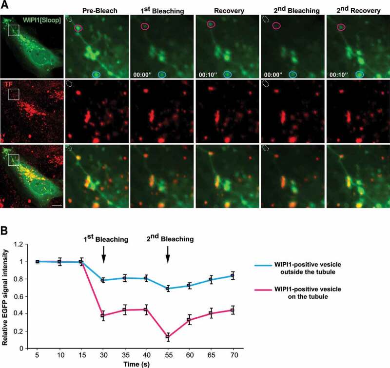
TF-enriched structures on WIPI1 tubules. (A) Photodepletion. HK2 cells were transfected with EGFP-WIPI1[Sloop]. After 18 h, they were loaded with Alexa Fluor 568-TF as in Figure 2 C. A region on a tubule (white dotted circle) was photobleached using a 488-nm laser. The images show concomitant photodepletion of EGFP-fluorescence from a TF-enriched punctate structure, which is outside the bleached area and associated with an EGFP-WIPI1[Sloop]-positive tubule. Two cycles of photodepletion and recovery were recorded. The boxed area of the cell in the image on the left is shown at higher magnification. Scale bar: 10 μm (B) Quantification of experiments as in (a). EGFP-fluorescence intensity was measured over time for EGFP-WIPI1[Sloop]-positive dots localized on a tubule (magenta circle in A), or far away from the tubule (light blue circle in A). Means ±s.d. are shown. 3 independent experiments with 10 cells each were analyzed