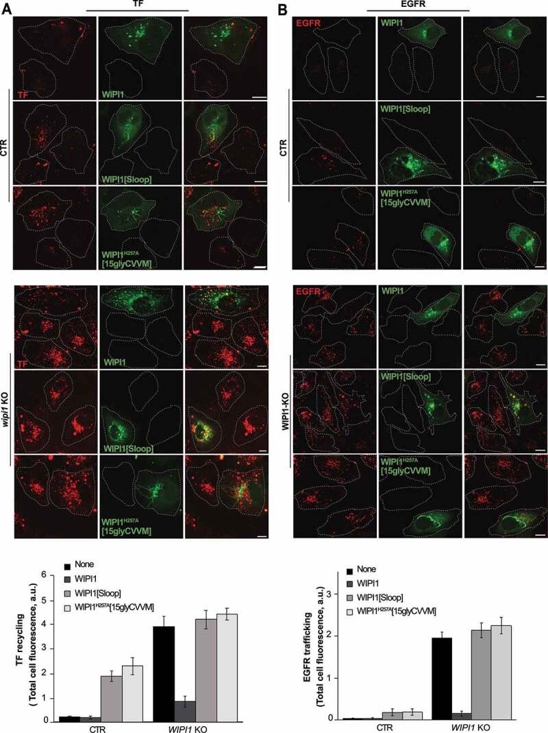
Figure 12.

Effect of WIPI1 mutants accumulating endosomal tubules on protein exit from endosomes. (A) TF recycling. Control and WIPI1 KO cells were transfected with EGFP-WIPI1, EGFP-WIPI1[Sloop] or EGFP-WIPI1H257A[15gly-CVVM] for 18 h. Then, they were serum-starved for 60 min, loaded with Alexa Fluor568-conjugated TF and chased at 37°C as in Figure 2 C. Scale bar: 10 μm. The white dashed lines indicate the circumference of the cells. TF-fluorescence was quantified as in Figure. 2B. Mean values ± s.d. are shown. n = 3 independent experiments with a total of 210 cells analyzed per condition. (B) EGFR degradation. CTR and WIPI1 KO cells expressing EGFP-WIPI1, EGFP-WIPI1[Sloop] and EGFP-WIPI1H257A[15gly-CVVM] as in (A) were serum-starved for 24 h and then stimulated with EGF (100 ng/ml) for 60 min. Cells were fixed, permeabilized, and stained with antibody to EGFR (red). Scale bar: 10 μm. EGFR-fluorescence was quantified as in Figure. 2B. 180 cells per condition stemming from 3 independent experiments were analyzed. Mean values ± s.d. are shown
