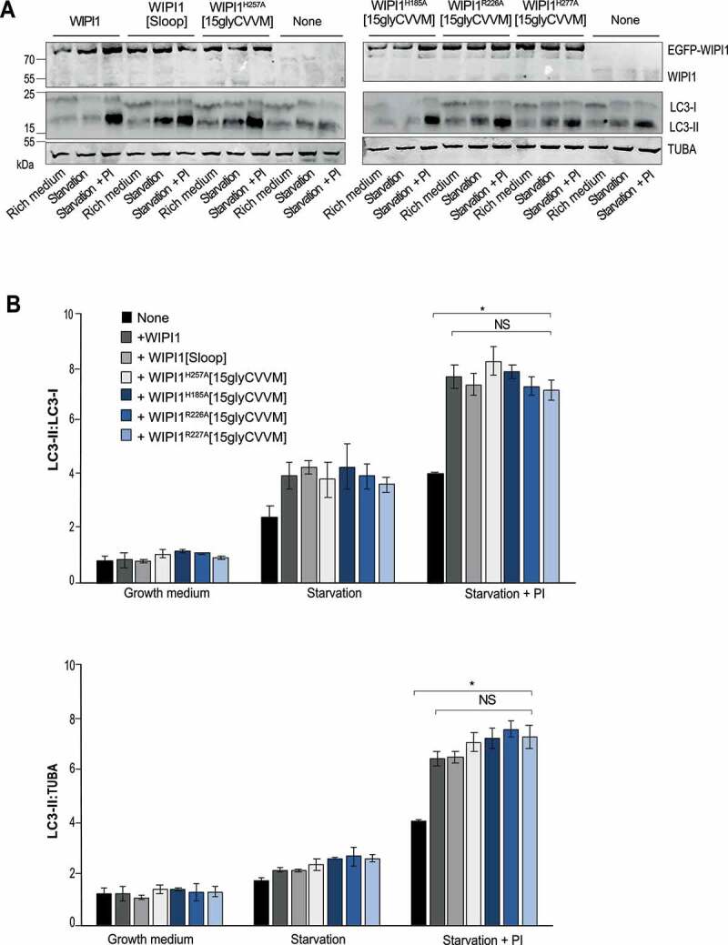
Figure 14.

Formation of LC3-II is not affected by WIPI1 mutations hindering protein exit from endosomes. (A) Total cell lysates of the cells from WIPI1 KO cells re-expressing the indicated WIPI1 variants (from Figure. 13 A; 50 μg of protein per sample) were analyzed by SDS–PAGE and western blotting using the indicated antibodies. TUBA/α-tubulin served as a loading control. A representative blot is shown. (B) The ratio LC3-II:LC3-I and LC3-II:TUBA/α-tubulin was quantified by a fluorescence scanner. Data are means ±s.d.; n = 3 independent experiments. P values are indicated and were calculated by t-Test. The analysis was performed with 99% confidence: *p < 0.01; NS = not significant)
