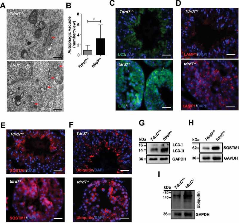
Figure 7.

Evaluation of Tdrd7+/− and tdrd7−/− seminiferous tubules. (A) Detection of Tdrd7+/− and tdrd7−/− mice testes using TEM. The red arrows indicate the autophagic vacuoles. (B) Quantification of the average number of autophagic vacuoles in Tdrd7+/− and tdrd7−/− mice testes using TEM analysis. *P < 0.05. (C-F) Immunostaining of Tdrd7+/− and tdrd7−/− seminiferous tubules for LC3 (C), LAMP1 (D), SQSTM1 (E), and ubiquitin (F) (red signal). The nuclei were stained with DAPI (blue signal). Scale bar: 50 μm. (G-I) Immunoblotting analysis of Tdrd7+/− and tdrd7−/− testicular tissue for LC3 (G), SQSTM1 (H), and ubiquitin (I) confirmed the accumulation of LC3-II, SQSTM1, and ubiquitin protein in tdrd7−/− seminiferous tubules
