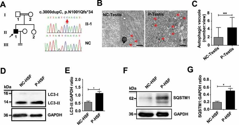
Figure 8.

An increase in autophagosomes was found in a patient with a TDRD7 mutation. (A) A homozygous frameshift mutation (c.3000dupC) of TDRD7 in a patient from a consanguineous family. The proband is indicated by the black arrow. The image on the right shows the sequence chromatograms for the proband and normal control (NC). The red arrow indicates the mutation site. (B) TEM detection of the seminiferous tubules in the patient with the TDRD7 mutation (c.324_325 insA) and NC. The red arrows indicate the autophagic vacuoles in the seminiferous tubules of the patient (P-testis) and NC. Scale bar: 2 μm. (C) Quantification of the average number of autophagic vacuoles in the seminiferous tubules of the patient and NC using TEM analysis. ***P < 0.001. (D-G) Western blotting detected the expression of LC3 (D) and SQSTM1 (F) in skin fibroblasts from the patient with a TDRD7 mutation (c.324_325 insA) and NC; GAPDH served as a loading control. Quantification analysis revealed significant accumulation of LC3-II (E) and SQSTM1 (G) (*P < 0.05). The data are representative of three independent assays
