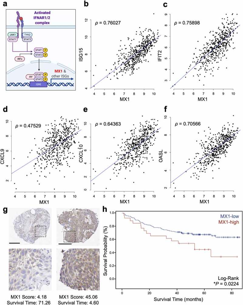
Figure 1.

High MX1 protein levels in cancer cells are a negative prognosticator in head and neck squamous cell carcinoma. (a) The signaling schematic shows that the IFNAR1 activation leads to the phosphorylation of STAT1/STAT2, which launches a transcriptional program centering on the ISGs, such as MX1. (b-f) Spearman correlation was performed between the expression levels of MX1 and representative ISGs including ISG15, IFIT2 (ISG54), CXCL9, CXCL10, and OASL using the HNSCC TCGA database (n = 520). (g) MX1 immunohistochemistry in TMAs from HNSCC patients reveals cytoplasmic staining of variable intensity across specimens. Raw MX1 scores and survival time are indicated for each representative TMA. Scale bar: 200 µm. (h) MX1 staining scores were segregated into MX1-low or MX1-high groups and the Kaplan-Meier survival curves of each group were compared using a log-rank test
