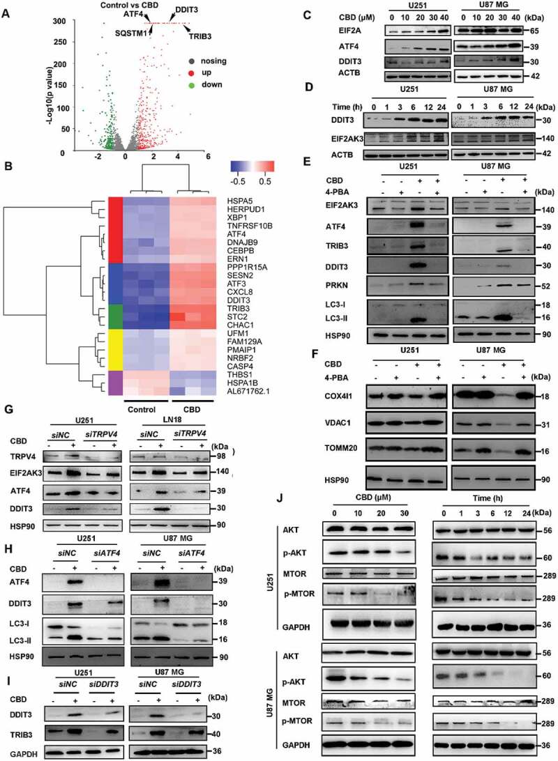
Figure 5.

CBD induces ER stress and mitophagy via the ATF4-DDIT3-TRIB3-AKT1-MTOR axis. (A) Transcriptome analysis of CBD-treated U251 cells. Volcano plot showing the up- and down-regulated genes, in red and green colors, respectively. Cells were treated with 30 μM CBD for 12 h. (B) Clustering analysis of stress-related genes in CBD-treated U251 cells. (C) Immunoblotting of ER stress markers EIF2A, DDIT3 and ATF4 in glioma cells after 24 h treatment with indicated concentrations of CBD. (D) Immunoblotting of DDIT3 and EIF2AK3 in glioma cells treated with 30 μM CBD for indicated times. (E-F) Effect of the ER stress inhibitor 4-PBA on CBD-induced mitophagy. Cells were treated with 20 μM CBD for 24 h with or without pretreatment with 4-PBA (1 mM for 30 min); expression levels of ER stress and mitophagy related proteins were analyzed by western blotting. (G) Effect of TRPV4 knockdown on CBD-induced ER stress. Cells with stable TRPV4 knockdown were treated with 30 μM CBD for 24 h, levels of TRPV4, EIF2AK3, ATF4 and DDIT3 were analyzed by western blotting. (H) Effect of ATF4 knockdown on CBD-induced ER stress and mitophagy. Cells were transfected with control or ATF4 siRNA for 48 h before treatment with CBD for another 24 h, express levels of ATF4, DDIT3, and LC3 were analyzed by western blotting. (I) Effect of DDIT3 knockdown on CBD-induced TRIB3 upregulation. Cells were transfected with control or DDIT3 siRNA for 48 h before treatment with CBD for another 24 h, express levels of DDIT3 and TRIB3 were analyzed by western blotting. (J) Effect of CBD treatment on the AKT-MTOR pathway. Cells were treated with CBD at indicated concentrations and times, expression levels of MTOR, p-MTOR, AKT and p-AKT were analyzed by western blotting. Immunoblots shown are representative of three independent experiments
