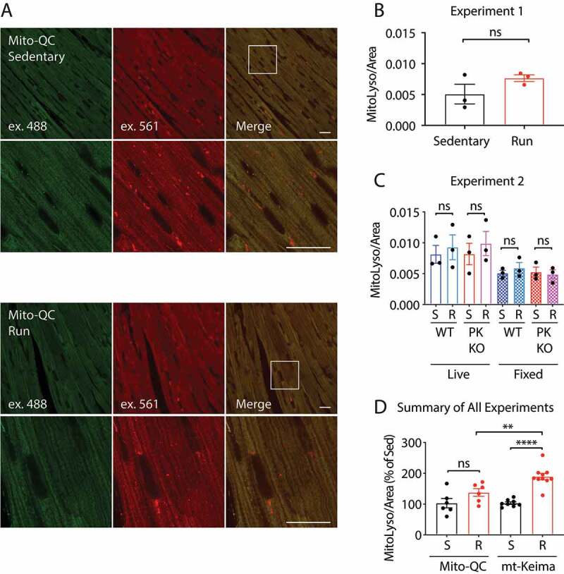
Figure 3.

Mito-QC does not detect PINK1-dependent mitophagy in vivo. (A and B) mito-QC mice were subjected to the protocol described in (Fig. 2A) and the tissue was immediately fixed in 4% paraformaldehyde and examined by confocal microscopy. Scale bar = 20 μm. mito-QC; pink1+/+ (WT) and mito-QC; pink1−/- (PK KO) littermates were either sedentary (S) or run (R), following the EE protocol in (Fig. 2A). For each animal a sample of heart tissue was immediately fixed and imaged later (fixed), and second sample was immediately imaged live (live). (D) Graph depicts sedentary (S) and run (R) data from all WT animals in experiments shown in Figure 2,Figure 3 normalized to sedentary animal mean values in each experiment. “ns” indicates not significant, “**” indicates p-value < 0.01, and “****” indicates p-value < 0.0001 after correction for multiple comparisons. Error bars represent standard error of the mean
