
Figure 3.
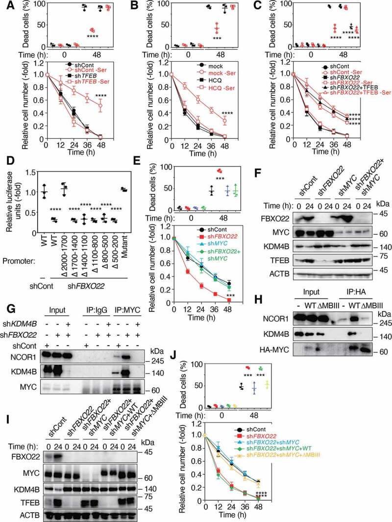
FBXO22 is required for proper TFEB transcription through the inhibition of MYC-KDM4B-NCOR1 transcription suppressor. (A) RPE-1 cells expressing the indicated Dox-inducible shRNA, preconditioned with or without serine depletion and then treated with low glucose (2 mM) media for 48 h. The proportions of dead cells (upper panel) and the relative cell numbers (lower panel) were determined at the indicated times as in Figure 1A. (B) RPE-1 cells were preconditioned as in (A) and then treated with low glucose (2 mM) media in the presence or absence of HCQ (10 μM). The proportions of dead cells (upper panel) and the relative cell numbers (lower panel) are determined at the indicated times. (C) RPE-1 cells expressing the indicated Dox-inducible shRNA and/or TFEB were preconditioned and treated as in (A). The proportions of dead cells (upper panel) and the relative cell numbers (lower panel) are determined at the indicated times. (D) HeLa cells expressing the indicated Dox-inducible shRNAs were transfected with the WT or the indicated deletion mutants of the TFEB promoter fused with the luciferase gene, and treated with doxycycline (1 μg/mL) for 48 h. The lysates were subjected to a luciferase assay. (E) RPE-1 cells expressing the indicated Dox-inducible shRNA, were preconditioned with serine depletion and then treated with low glucose (2 mM) media for 48 h. The proportions of dead cells (upper panel) and the relative cell numbers (lower panel) were determined at the indicated times as in (A). (F) Lysates from RPE-1 cells as in (E) at the indicated times after incubation with low glucose media were subjected to immunoblotting using the indicated antibodies. (G) Lysates from HeLa cells expressing the indicated Dox-inducible shRNAs were immunoprecipitated and then subjected to immunoblotting using the indicated antibodies. (H) Lysates from RPE-1 cells expressing WT or a MYC Box III deletion mutant (ΔMBIII) MYC were immunoprecipitated and then subjected to immunoblotting using the indicated antibodies. (I) Lysates from RPE-1 cells expressing the indicated Dox-inducible shRNAs and/or WT MYC or ΔMBIII at the indicated times after serine depletion were subjected to immunoblotting using the indicated antibodies. (J) Cells as in (I) were preconditioned with serine depletion and then treated with low glucose (2 mM) media for 48 h. The proportions of dead cells (upper panel) and the relative cell numbers (lower panel) were determined. Data are presented as means±s.d. of three independent experiments. One-way ANOVA with Dunnett’s multiple comparisons post hoc test was performed against a control (shCont or mock) (A-E, J). ***P < 0.001, ****P < 0.0001