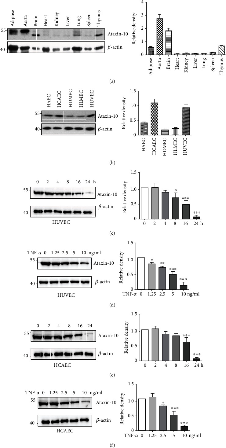
Figure 1.

Expression of Ataxin-10 in endothelial cells. (a) Representative images of Western blotting for detection of the expression of Ataxin-10 in mouse tissues including adipose, aorta, brain, heart, kidney, liver, lung, spleen, and thymus. β-Actin was provided as a loading control. (b) Representative images of Western blotting for detection of the expression of Ataxin-10 in human primary vascular endothelial cells. (c, d) HUVEC or (e, f) HCAEC was stimulated with TNF-α for different time intervals and doses as indicated. Cell lysates were collected for Western blotting analysis. Quantification of the bands was carried out using Gel-Pro Analyzer software, and results were presented as “fold changes” at the right side of the bands.
