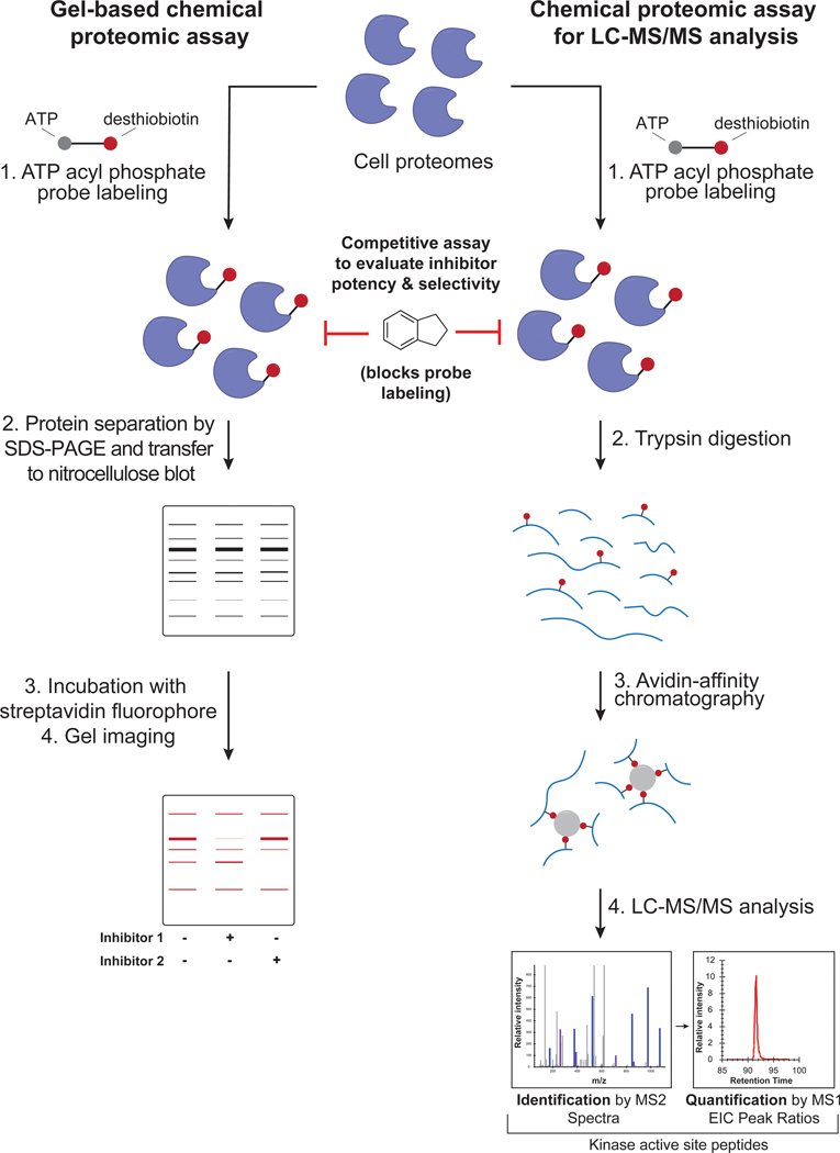
Figure 2. Workflow of gel- and LC-MS/MS-based chemical proteomic assay.
Left: schematic of gel-based chemical proteomic assay. Cell lysates are incubated with ATP acyl phosphate probe, and the probe-labeled proteins separated by SDS-PAGE and then transferred to a nitrocellulose blot. The blot is incubated with a streptavidin fluorophore and then imaged to reveal probe-labeled protein bands. Right: schematic of chemical proteomic assay for LC-MS/MS analysis. Cell lysates are incubated with ATP acyl phosphate probe, proteins digested into peptides with trypsin, and probe-modified peptides enriched using avidin resin. The resulting peptides are analyzed by LC-MS/MS.

