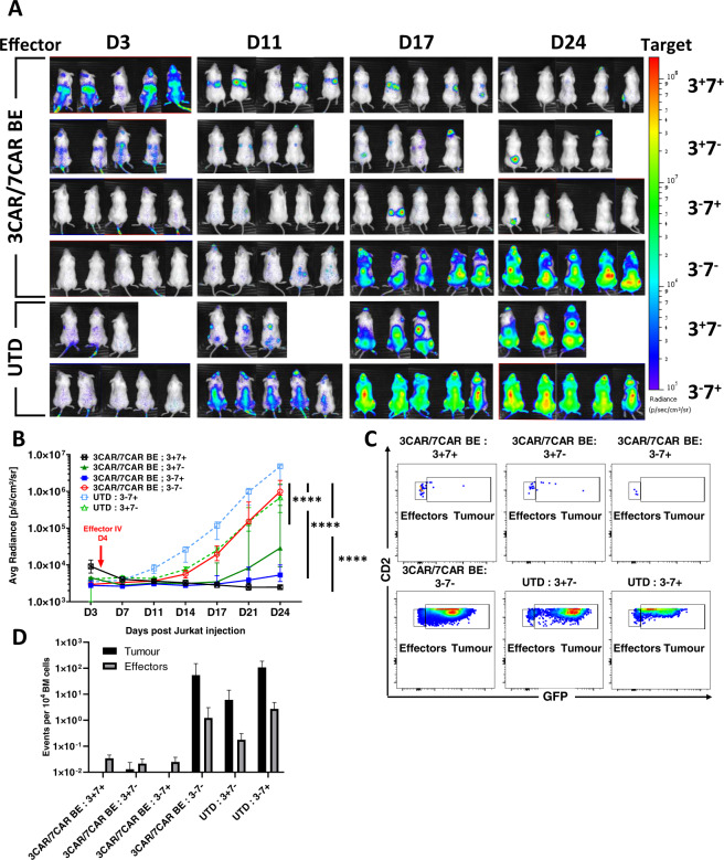
Fig. 6. 3CAR and 7CAR cells effectively clear T cell malignancy in vivo.
A NSG mice were infused with 1 × 10⁷ GFP + LUC + Jurkat T cells modified to express mixed CD3 and/or CD7 surface antigens in groups of (n = 5) CD3−CD7−, (n = 4) CD3+CD7−, (n = 5) CD3−CD7+ or (n = 5) CD3−CD7− and imaged on day 3 prior to infusion of 1 × 10⁷ TCR−CD7− 3CAR/7CAR mixed effectors or untransduced (UTD) cells. Leukaemic progression monitored by serial BLI for 24 days and revealed disease progression in animals receiving untransduced T cells (3CAR−7CAR−) and in animals engrafted with antigen-negative (CD3−CD7−) leukaemia. B Bioluminescence signal of each animal plotted as Average radiance [photons/s/cm2/sr]. Each line represents a different experimental group and each point on the line the mean of each group. Error bars represent SEM. Area under the curve was calculated for each experimental group and values were compared using a one-way ANOVA with Tukey multiple comparison post-hoc****P < 0.0001. C Example of day 24 flow cytometry-based detection in bone marrow of mCD11b−/hCD45+ effector T cells (CD2+GFP−) in 3CAR/7CAR treated animals and residual leukaemia (CD2+GFP+) in antigen-negative and untransduced groups. D Frequency of hCD45+CD2+GFP− effector events or hCD45+CD2+GFP+ Jurkat events per 104 acquired bone marrow events.

