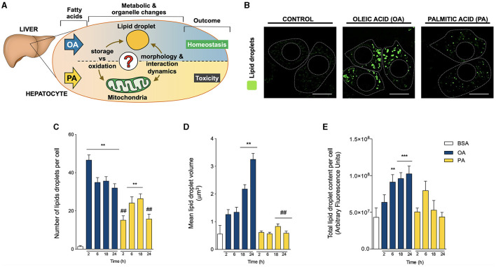
Figure 1.
Oleic and palmitic acids differentially remodel lipid droplet morphology in HepG2 cells. (A) In the liver, oleic acid (OA) overload seemingly does not damage hepatocyte function, while palmitic acid (PA) is evidently toxic. To date, the underlying mechanism of this difference remains unknown in terms of fatty acid metabolism and mitochondria-lipid droplet interactions and morphology. (B) Representative confocal imaging of HepG2 cells treated with 200 μM of OA or PA during 18 h and stained with Bodipy 493/503 (green) for lipid droplet (LD) visualization. Segmented lines represent the cellular and nuclear contours. Scale bar: 10 μm. (C) Quantification of the number of LDs per cell in images treated as in A for 0–24 h. (D) Mean individual volume of LDs in HepG2 cells treated as in A for 0–24 h. (E) Quantification of the total Bodipy 493/503 fluorescence in HepG2 cells treated as in A for 0–24 h. Data are from N = 6–20 cells analyzed from three independent experiments. Results are shown as mean ± SEM; *p < 0.05; **p < 0.01; ***p < 0.001 vs. BSA; # p < 0.05; and ## p < 0.01 vs. OA at the same time conditions.

