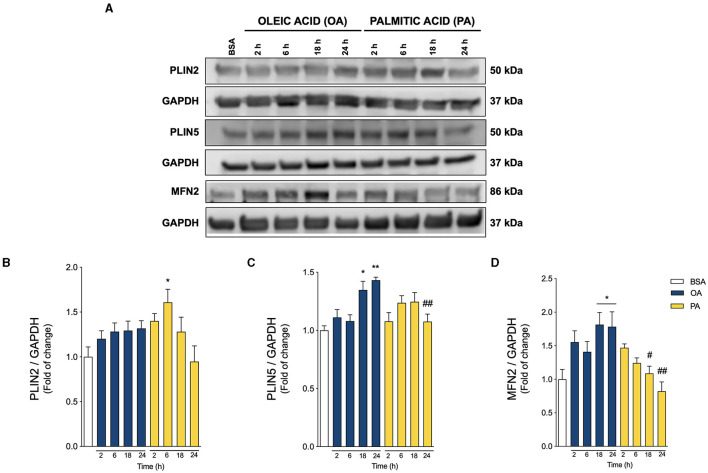
Figure 3.
Oleic and palmitic acids induce different patterns of lipid droplet-mitochondria tether proteins in HepG2 cells. (A) Representative Western blots of the proteins associated with lipid droplet (LD)-mitochondria physical interaction: Perilipin 2 and 5 (PLIN2, PLIN5), and Mitofusin 2 (MFN2) in HepG2 cells treated with 200 μM of oleic acid (OA) or palmitic acid (PA) for 0–24 h. GAPDH was used as a loading control. (B–D) Densitometric quantification of the proteins indicated in A. Data are expressed as mean ± SEM of N = 7 for PLIN2 and N = 4 for PLIN5 and MFN2. * p < 0.05; ** p < 0.01 vs. BSA; # p < 0.05; and ## p < 0.01 vs. OA at the same time conditions.

